Tresserra F, Fabra G, Luque O, Castélla M, Gómez C, Fernández-Cid C, Rodríguez I.
Rev Esp Patol. 2024 Jul-Sep;57(3):182-189 . DOI: 10.1016/j.patol.2024.03.005
The difficulty in examining cytological smears lies in the surface area they occupy and the three-dimensionality of certain cell groups. Our centre has recently implemented a digital imaging system for liquid cytology that combines a novel artificial intelligence (AI) algorithm with advanced volumetric imaging technology. The use of liquid-based cytology allows for a homogeneous, single-layer study area, minimising observation problems.
While numerous histology guidelines provide recommendations for the validation of digital systems, there are still no clear references in cytology. Histology guidelines recommend a minimum of 60 representative cases per practitioner over a two-week period with a minimum concordance of 95% between techniques.
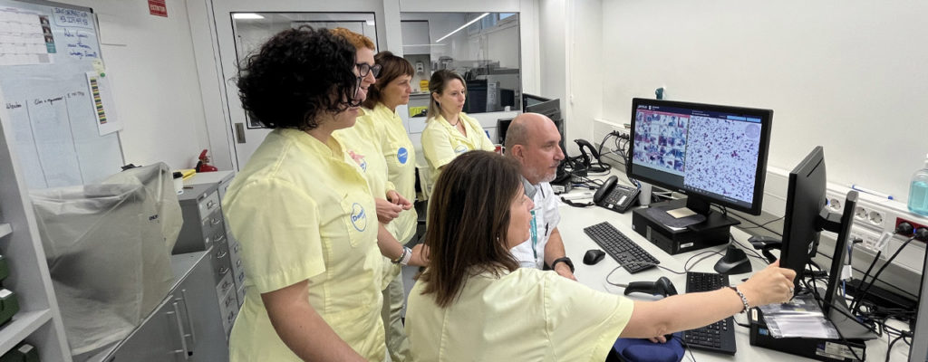
Following these recommendations, and with the aim of testing the diagnostic efficiency of digital systems compared to conventional microscopy, a team of researchers from the Cervical Pathology Unit of Dexeus Mujer and the Hospital Universitari Dexeus, led by Dr Francesc Tresserra, conducted a specific validation study. The team, consisting of five cytotechnologists (CT), reviewed 888 routine cervico-vaginal (CV) cytology cases from the Cervical Pathology Unit of our centre over a two-week period. Cases were initially observed by microscopy and later by digital imaging, with concordance measured using the kappa index.
According to the authors, the results showed a high concordance between the two systems, confirming the diagnostic validity of the digital technique in cervico-vaginal cytology.